
Applied Microscopy and Cell Biology
The laboratory is actively involved in the development of new protocols and advanced cellular models for biocompatibility, toxicity and regenerative medicine studies, exploiting cutting-edge technologies.
Activities
Cellular Models to evaluate Compounds Biocompatibility and Toxicity :
- BIOLOGICAL SAFETY ASSESSMENT & BIOCOMPATIBILITY TESTING according to ISO10993-5, ISO10993-4, ISO10993-23 and in vitro sensitization test according to OECD442D and OECD442E.
- DESIGN AND DEVELOPMENT OF ADVANCED BIOLOGICAL MODELS FOR BIOCOMPATIBILITY STUDIES: 2D cultures or Bioprinting are applied for the development of customized models to investigate the efficacy and safety of drugs and medical device, in order to highlight performance and critical aspect reducing time to market. MAB lab has skills in cell culture isolation form healthy and pathological tissues and know-how on isolation, characterization and culture of mesenchymal stroma cells from multiples tissue and species.
- INTERACTION CELL-MATERIAL: 3D cell cultures are performed on biomaterials and medical device to evaluate citotoxicity and biocompatibility. The dual interaction between cells and biomaterials among the development of specific tissue is investigate thanks to advanced microscopic technologies
- BIOPRINTING: In collaboration with the MS2 laboratory, it is possible to print both cells and materials to obtain innovative cellular and tissue models able to mimic the physiology of organs and tissues. The ultimate goal is to predict safety, biocompatibility and effectiveness of new therapeutic approaches.
- ADVANCED CELL THERAPIES AND REGENERATIVE MEDICINE
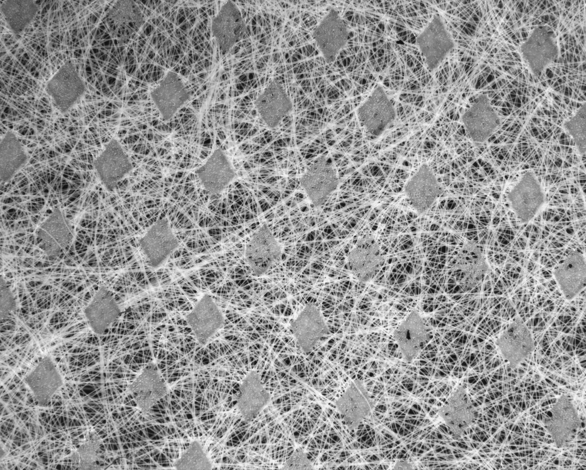
- PARTICLE COUNT ON FILTER
- MOLECOULAR BIOLOGY INVESTIGATIONS: Gene Expression evaluation by real-time PCR, acid nucleic extraction from cell cultures, tissues and 3D cell cultures
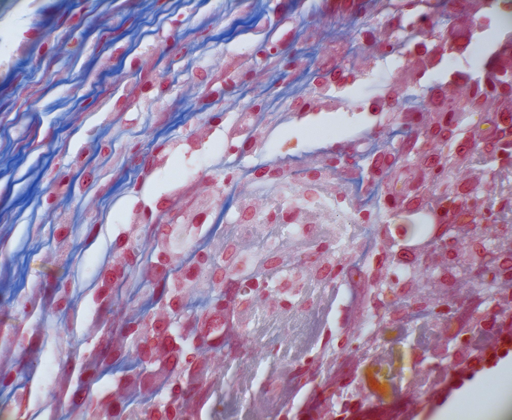
- HISTOLOGY INVESTIGATIONS: the lab is equipped for paraffin embedding of biological specimens, paraffin section staining, immunohistochemistry and immunofluorescence techniques.
- SCIENTIFIC MANUSCRIPTS, ETICHAL COMMIETTES AND PATENTES
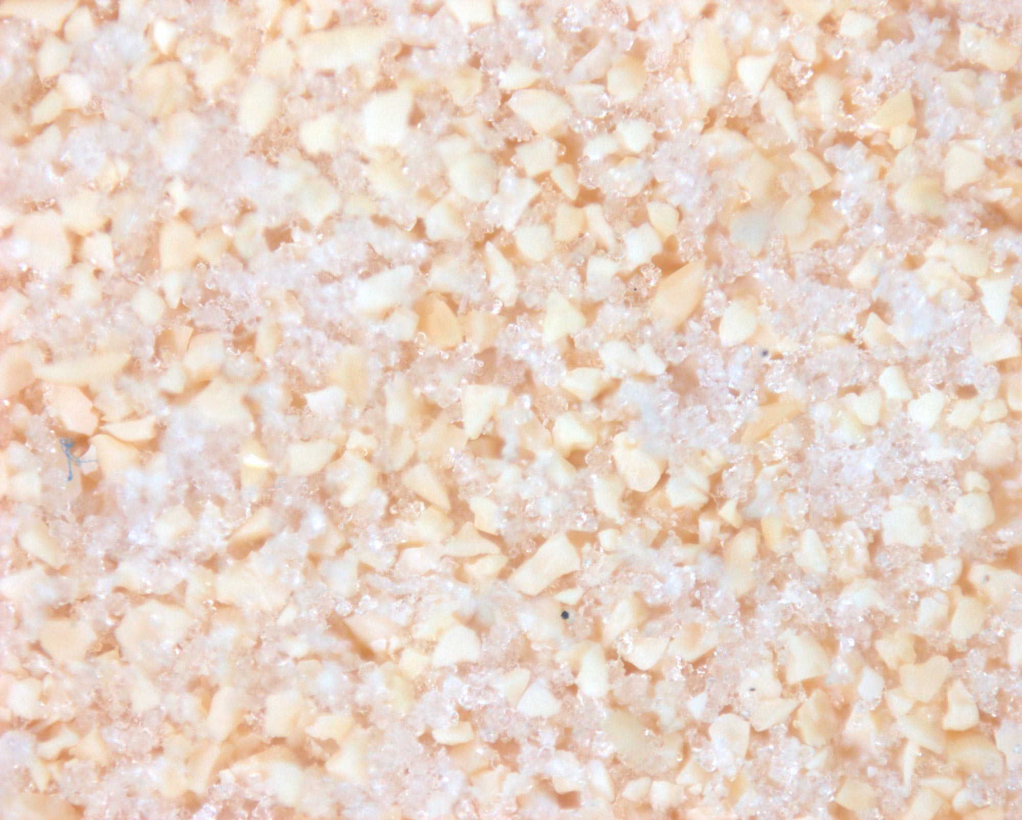
SURFACE CHARACTERIZATION AND PARTICLE COUNT: staining and advanced optical microscopic combined with electronic microscopic (SEM), IR and chemistry interface (XPS) are applied to investigate the material surface - DETECTION OF BLOOD CELLS AND/OR BIOFILM ON A MATERIAL: histology approaches are applied to a medical device to characterize the distribution of blood cells and clots or bacteria biofilm in a medical device after ex-vivo use to prevent critical aspects during clinical application
Application sectors
Dental
Pharmaceutical
Cosmetic
Agro-food
Biomedical
Regenerative Medicine:
Identification of the cell-biomaterial combination, which allows the differentiation towards a target tissue.
MAB has experience in:
- pre-clinical studies for the regeneration of bone and adipose tissue
- Writing and submission of studies to the ethics committee for in vivo experiments
- In vivo damage simulation of human pathologies and treatments with stem cells and biomaterials in autologous, allogeneic and xenogenic settings
- assessment of the implant through histological investigations
Application sectors
Pharmaceutical
Veterinary
Microscopy, Image Analysis, Histology:
The advanced microscopy branch of the MAB laboratory is able to answer to different company needs through :
- histopathological investigations for the physio-pathological characterization of tissues
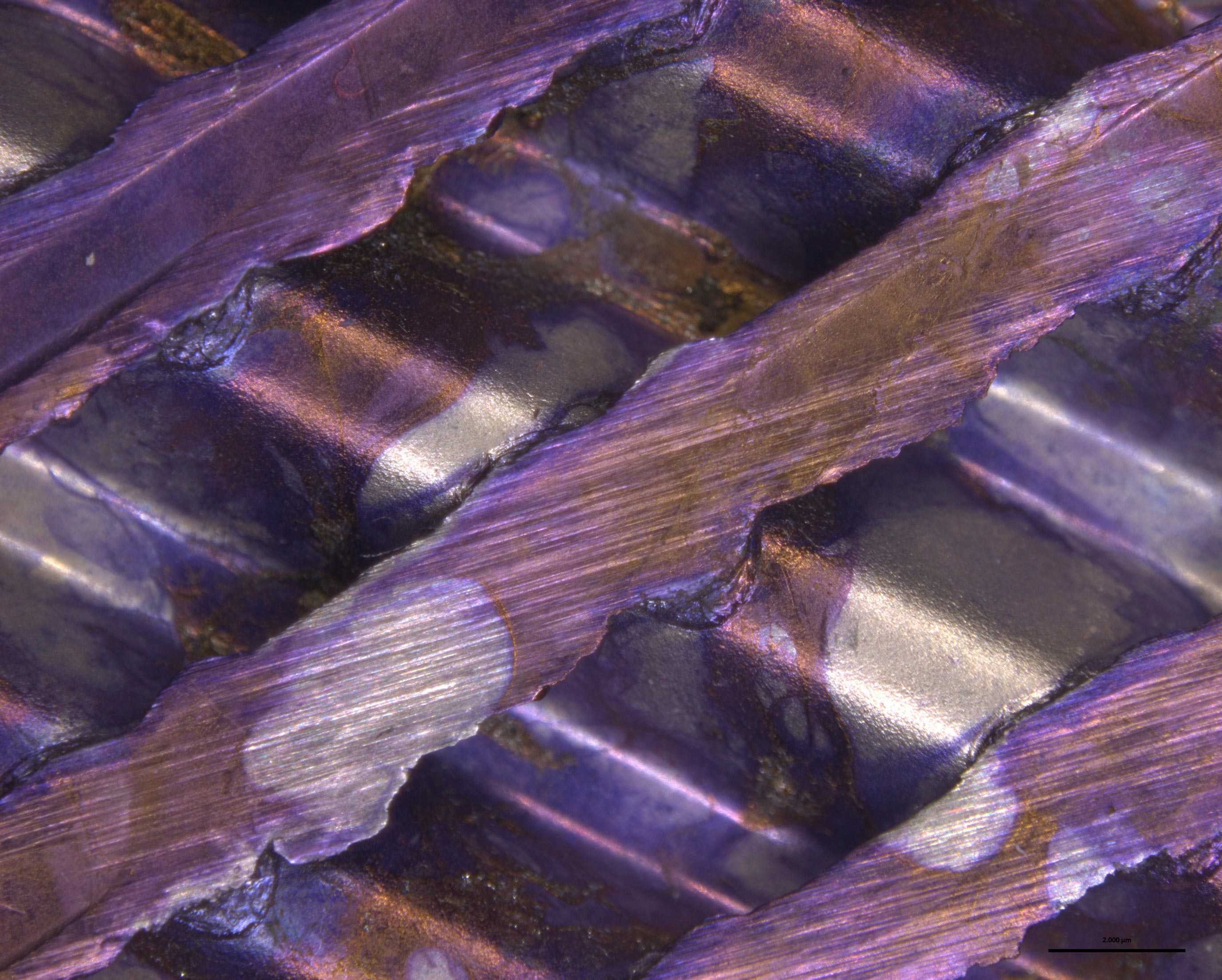
- surface studies of materials by large surfaces scanning with high resolving power and reconstruction of the image in three dimensions
- biomaterials colonization with fluorescent cells
- image analysis to transform qualitative data into a semi-quantitative survey
We deliver tailor made approaches through:
- creation of three-dimensional models of bacterial biofilms on biomedical devices for the evaluation of sterilization processes or new functionalizations of surfaces, in vivo and in vitro.
- biochemical tests for the study of inflammatory processes in various stages of treatment.
- blood simulation models to detect possible blood parameters changes in and activation of coagulation processes.
- functionalization and characterization of membrane performances for biomedical devices.
- support in the R & D phases.
Application sectors
Cosmetic
Agro-Food
Veterinary
Analysis of bacterial biofilms on biomedical devices surfaces :
It is possible to evaluate the bacterial biofilms formation on a biomedical device through in vitro and in vivo investigations, monitoring the same animal with in vivo imaging. Microscopy techniques can also be applied to study the biofilm distribution in the device.
Application sectors
Biomedical
Ceramic-Building
Scientific director
Prof. Massimo Dominici MD
Laboratory manager
Dott. Elena Veronesi PhD
Staff and skills
Dott. Elena Veronesi: Laurea in Scienze Biologiche, PhD in Medicina Molecolare e Rigenerativa
Dott. Elisa Resca: Laurea in Biotecnologie Mediche, PhD in Scienze Morfologiche Umane e Molecolari
Dott. Tiziana Petrachi: Laurea in Biotecnologie Mediche e Farmaceutiche, PhD in Medicina Molecolare e Rigenerativa
Dott. Valentina Bergamini: Laurea in Biotecnologie Mediche, esperta di colture di cellule staminali, PhD student
Dott. Francesco Ganzerli: Laurea in Biotecnologie Mediche, esperto di colture di cellule staminali e skin equivalent

Instrumentation
Axiozoom V16 (Zeiss) optical microscope that combines both stereomicroscope and confocal features. Versatile tool, suitable for many applications, for surface observation of plastics, organic materials, metals, cements, biological material adhered to materials, cell cultures.
Enspire multi-plate reader (PerkinElmer) for evaluation of cytokines released following stimulation, evaluation of dye concentrations, label free assays, receptor activation studies.
Instrumentation for cell cultures: cell cultures hoods, incubators, centrifuge, bath thermostat, tissue digestion shaker, dedicated refrigerators, inverted optical microscope (Observer, Zeiss) and EVOS (life technologies), freezer -80 and nitrogen plant for cell preservation.
Station for paraffin embedding: automatic inclusor, microtome, fluorescence-equipped optical microscope (Imager, Zeiss)